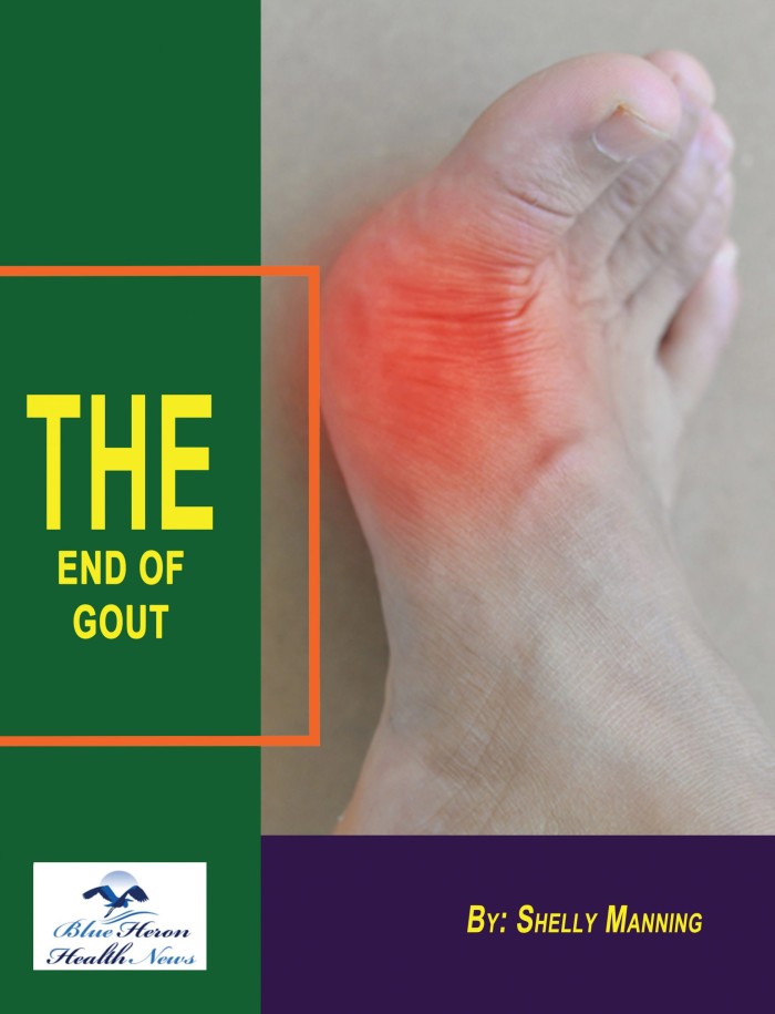
The End Of GOUT Program™ By Shelly Manning The program, End of Gout, provides a diet set up to handle your gout. It is a therapy regimen for gout sufferers. It incorporates the most efficient techniques and approaches to be implemented in your daily life to heal and control gout through the source.
How can one differentiate between gout and pseudogout?
Differentiating between gout and pseudogout can be challenging because both conditions involve joint inflammation and share similar symptoms. However, they are distinct diseases caused by different types of crystal deposits in the joints. Here is a comprehensive guide to understanding the differences between gout and pseudogout, including their causes, symptoms, diagnostic procedures, and treatment options:
1. Causes and Pathophysiology
Gout
- Crystals Involved: Gout is caused by the deposition of monosodium urate (MSU) crystals in the joints.
- Uric Acid Levels: Elevated levels of uric acid in the blood (hyperuricemia) lead to the formation of these crystals.
- Risk Factors: Risk factors for gout include genetics, diet high in purines (red meat, shellfish), alcohol consumption, obesity, hypertension, chronic kidney disease, and certain medications (e.g., diuretics).
Pseudogout
- Crystals Involved: Pseudogout, also known as calcium pyrophosphate deposition disease (CPPD), is caused by the deposition of calcium pyrophosphate dihydrate (CPP) crystals in the joints.
- Calcium Metabolism: Issues with calcium metabolism or cartilage damage can contribute to CPPD. The exact cause is not always clear.
- Risk Factors: Risk factors for pseudogout include aging, joint trauma, genetic factors, metabolic disorders (e.g., hyperparathyroidism, hemochromatosis), and osteoarthritis.
2. Symptoms
Common Symptoms
- Joint Pain: Both conditions cause sudden, severe joint pain.
- Swelling and Redness: The affected joint may become swollen, red, and warm to the touch.
- Limited Mobility: Joint stiffness and reduced range of motion are common.
Gout-Specific Symptoms
- Affected Joints: Gout often affects the big toe (podagra) but can also involve other joints like the ankles, knees, elbows, wrists, and fingers.
- Tophi: Chronic gout can lead to the formation of tophi, which are lumps of urate crystals under the skin around the joints.
- Attack Pattern: Gout attacks can be sudden and severe, often occurring at night and lasting from a few days to a week. Intervals between attacks can vary.
Pseudogout-Specific Symptoms
- Affected Joints: Pseudogout commonly affects larger joints such as the knees, wrists, shoulders, and elbows.
- Chronic Joint Pain: Unlike gout, pseudogout can present with chronic joint pain and intermittent flare-ups.
- Attack Pattern: Pseudogout attacks can be less sudden and severe compared to gout but can still cause significant pain and discomfort.
3. Diagnostic Procedures
Medical History and Physical Examination
- Symptom Review: A detailed medical history and review of symptoms are essential. The pattern of joint involvement and the nature of the attacks can provide clues.
- Physical Exam: A physical examination to assess joint swelling, redness, warmth, and tophi (in gout) can help differentiate the conditions.
Laboratory Tests
- Serum Uric Acid: Elevated serum uric acid levels are indicative of gout. However, normal levels do not rule out gout.
- Serum Calcium and Phosphate: Abnormal calcium and phosphate levels can suggest pseudogout, especially in the context of metabolic disorders.
Synovial Fluid Analysis
- Joint Aspiration: Aspiration of synovial fluid from the affected joint is a key diagnostic procedure for both conditions.
- Crystal Identification: Under a polarizing microscope:
- MSU crystals (gout) appear needle-shaped and show strong negative birefringence (yellow when aligned parallel to the axis of the compensator).
- CPP crystals (pseudogout) appear rhomboid or rod-shaped and show weak positive birefringence (blue when aligned parallel to the axis of the compensator).
Imaging Studies
- X-Rays: Can reveal joint damage and the presence of tophi in chronic gout or chondrocalcinosis (calcium deposits in cartilage) in pseudogout.
- Ultrasound: Can detect urate crystal deposits in gout and CPP deposits in pseudogout.
- Dual-Energy CT (DECT): Highly effective in identifying urate crystals in gout, but less commonly used for pseudogout.
4. Treatment Options
Acute Attack Management
- NSAIDs: Nonsteroidal anti-inflammatory drugs (NSAIDs) are commonly used to relieve pain and inflammation in both conditions.
- Corticosteroids: Oral or intra-articular corticosteroids can be used to manage acute flare-ups of both gout and pseudogout.
- Colchicine: Effective in treating acute gout attacks and can also be used in pseudogout, though it may be less effective.
Long-Term Management
- Gout
- Urate-Lowering Therapy: Medications like allopurinol or febuxostat to lower uric acid levels and prevent future attacks.
- Lifestyle Modifications: Dietary changes to reduce purine intake, alcohol reduction, weight management, and regular exercise.
- Regular Monitoring: Regular monitoring of uric acid levels to ensure they remain within target ranges.
- Pseudogout
- Management of Underlying Conditions: Treating underlying metabolic disorders (e.g., hyperparathyroidism) that contribute to CPPD.
- Joint Health Maintenance: Physical therapy and exercises to maintain joint function and mobility.
- Periodic Joint Aspiration: In cases of recurrent effusions, periodic joint aspiration may be necessary.
5. Prognosis and Follow-Up
Gout
- Prognosis: With proper management, most patients with gout can achieve good control of their symptoms and prevent joint damage.
- Follow-Up: Regular follow-up with a healthcare provider to monitor uric acid levels and adjust treatment as necessary.
Pseudogout
- Prognosis: While there is no cure for pseudogout, effective management of symptoms and underlying conditions can improve quality of life and prevent complications.
- Follow-Up: Regular follow-up to manage chronic symptoms and monitor for joint health.
Conclusion
Differentiating between gout and pseudogout involves understanding the underlying causes, symptoms, diagnostic procedures, and treatment options for each condition. Accurate diagnosis through detailed medical history, physical examination, laboratory tests, synovial fluid analysis, and imaging studies is crucial. Early and appropriate treatment can significantly improve outcomes and quality of life for individuals with either condition. Regular follow-up and management of underlying risk factors are essential to prevent complications and maintain joint health.

The End Of GOUT Program™ By Shelly Manning The program, End of Gout, provides a diet set up to handle your gout. It is a therapy regimen for gout sufferers. It incorporates the most efficient techniques and approaches to be implemented in your daily life to heal and control gout through the source.