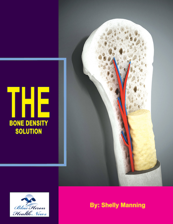
The Bone Density Solution by Shelly Manning As stated earlier, it is an eBook that discusses natural ways to help your osteoporosis. Once you develop this problem, you might find it difficult to lead a normal life due to the inflammation and pain in your body. The disease makes life difficult for many.
What is a DEXA scan?
Comprehensive Guide to DEXA Scan
A DEXA scan, or Dual-Energy X-ray Absorptiometry, is a medical imaging technique used to measure bone density. It is considered the gold standard for diagnosing osteoporosis and assessing fracture risk. This comprehensive guide explores the technology, procedure, clinical applications, interpretation of results, benefits, risks, and advancements in DEXA scanning.
1. Introduction to DEXA Scan
1.1 Definition
- DEXA Scan: A non-invasive imaging test that uses low-dose X-rays to measure bone mineral density (BMD). It helps diagnose conditions like osteoporosis and assess the risk of bone fractures.
1.2 History
- Development: DEXA technology was developed in the 1980s, building on earlier methods like single-photon and dual-photon absorptiometry. It became widely used in the 1990s due to its accuracy and reliability.
- Evolution: Over the years, advancements in technology have improved the precision and application of DEXA scans.
2. Technology Behind DEXA Scan
2.1 How It Works
- X-ray Technology: DEXA uses two X-ray beams at different energy levels. One beam is high energy, and the other is low energy.
- Bone Density Measurement: The amount of X-ray energy absorbed by the bones and soft tissues is measured. The difference between the two energy levels helps calculate bone density.
2.2 Equipment
- Scanning Device: A DEXA machine consists of a flat, padded table where the patient lies, and an overhead scanning arm that moves over the body.
- Detector: The detector measures the X-rays that pass through the body, providing data on bone density and composition.
2.3 Radiation Exposure
- Low Dose: The radiation exposure from a DEXA scan is minimal, comparable to natural background radiation received over a day.
3. Procedure for DEXA Scan
3.1 Preparation
- Patient Preparation: Minimal preparation is needed. Patients may be advised to avoid calcium supplements for 24 hours before the test.
- Clothing: Comfortable clothing without metal fasteners is recommended. Patients may need to change into a gown.
3.2 Scan Process
- Positioning: The patient lies on the DEXA table, usually on their back. The technician positions the patient to ensure accurate scanning of the spine, hip, or forearm.
- Scanning: The scanning arm passes over the body, emitting X-rays and capturing images. The process is quick, typically taking 10-20 minutes.
- Comfort: The procedure is painless, and patients are generally comfortable throughout the scan.
4. Clinical Applications of DEXA Scan
4.1 Diagnosing Osteoporosis
- Bone Density Measurement: DEXA is primarily used to measure bone density in the spine, hip, and sometimes the forearm.
- T-Score: Results are expressed as T-scores, comparing the patient’s bone density to that of a healthy young adult. A T-score of -2.5 or lower indicates osteoporosis.
4.2 Assessing Fracture Risk
- Risk Prediction: DEXA scans help predict the risk of fractures, particularly in postmenopausal women and older adults.
- FRAX Tool: The Fracture Risk Assessment Tool (FRAX) incorporates DEXA results with other risk factors to estimate the 10-year probability of fractures.
4.3 Monitoring Bone Health
- Treatment Monitoring: Regular DEXA scans are used to monitor changes in bone density over time, assessing the effectiveness of treatments for osteoporosis and other bone-related conditions.
4.4 Other Applications
- Body Composition Analysis: Advanced DEXA scans can provide detailed information on body composition, including the distribution of fat and lean tissue.
- Research Studies: DEXA is used in clinical research to study bone health, aging, and the effects of various treatments on bone density.
5. Interpretation of DEXA Scan Results
5.1 T-Score and Z-Score
- T-Score: Compares the patient’s bone density to that of a healthy young adult. Scores are classified as normal (-1.0 or above), osteopenia (-1.0 to -2.5), and osteoporosis (-2.5 or below).
- Z-Score: Compares the patient’s bone density to an age-matched reference population. A Z-score below -2.0 may indicate secondary causes of bone loss.
5.2 Comprehensive Report
- Detailed Analysis: The DEXA scan report includes images, T-scores, Z-scores, and a summary of findings.
- Clinical Recommendations: Based on the results, healthcare providers may recommend lifestyle changes, medications, or further tests.
6. Benefits of DEXA Scan
6.1 Accuracy and Precision
- High Accuracy: DEXA scans provide precise measurements of bone density, essential for diagnosing and managing osteoporosis.
- Reliability: The method is reliable, with minimal variability in repeated measurements.
6.2 Non-Invasive and Quick
- Painless Procedure: The scan is non-invasive and painless, making it suitable for regular monitoring.
- Quick Process: The procedure is fast, usually completed within 10-20 minutes.
6.3 Low Radiation Exposure
- Minimal Risk: The low dose of radiation makes DEXA scans safe for repeated use, essential for monitoring bone health over time.
7. Risks and Limitations of DEXA Scan
7.1 Radiation Exposure
- Low Radiation Risk: While the radiation dose is low, it is still present. Pregnant women should inform their healthcare provider before undergoing a DEXA scan.
- Cumulative Exposure: Repeated scans over time can lead to cumulative radiation exposure, though it remains minimal compared to other imaging techniques.
7.2 Limitations in Measurement
- Site Specificity: DEXA scans primarily measure bone density in specific areas like the spine and hip, which may not reflect bone density throughout the entire skeleton.
- Body Composition Interference: High levels of fat or other tissue can sometimes interfere with the accuracy of bone density measurements.
7.3 False Results
- Artifacts: Artifacts such as surgical implants, calcified arteries, or previous fractures can affect the accuracy of the scan.
- Calibration Issues: Variations in machine calibration can lead to discrepancies in results, though this is rare with modern equipment.
8. Advancements in DEXA Technology
8.1 Enhanced Imaging Techniques
- 3D Imaging: Advanced DEXA machines provide three-dimensional imaging for more detailed assessment of bone structure.
- High-Resolution Scans: Improvements in resolution offer better visualization of bone microarchitecture.
8.2 Software Improvements
- Body Composition Analysis: New software allows for detailed analysis of body composition, including fat distribution and muscle mass.
- Automated Reporting: Enhanced software capabilities provide automated, comprehensive reports with clinical recommendations.
8.3 Integration with Other Diagnostic Tools
- Combined Assessments: Integration with other diagnostic tools like MRI and CT scans offers a more comprehensive evaluation of bone health.
- Research Applications: Ongoing research explores the use of DEXA in new areas, such as cardiovascular health and metabolic disorders.
9. Case Studies and Personal Stories
9.1 Clinical Case Studies
- Diagnosis and Treatment: Examples of patients diagnosed with osteoporosis through DEXA scans and their subsequent treatment plans.
- Monitoring Progress: Case studies showing how regular DEXA scans helped monitor treatment efficacy and adjust therapeutic strategies.
9.2 Personal Stories
- Patient Experiences: Real-life stories of individuals undergoing DEXA scans, their diagnosis, and their journey in managing bone health.
10. Public Health and Awareness
10.1 Education and Outreach
- Awareness Campaigns: Public health initiatives to educate the public about the importance of bone density measurement and osteoporosis prevention.
- Community Programs: Local programs offering DEXA scans, educational workshops, and resources for maintaining bone health.
10.2 Policy and Advocacy
- Healthcare Policies: Advocacy for policies supporting access to bone density testing and osteoporosis treatment.
- Support Networks: Building support networks for individuals diagnosed with osteoporosis to provide information, resources, and emotional support.
11. Conclusion
A DEXA scan is a vital tool for measuring bone density, diagnosing osteoporosis, assessing fracture risk, and monitoring bone health. With its high accuracy, low radiation exposure, and non-invasive nature, DEXA remains the gold standard for bone density measurement. Advancements in technology continue to enhance its capabilities, offering more detailed assessments and better integration with other diagnostic tools. Public health initiatives and patient education are crucial for promoting awareness and ensuring that individuals at risk of osteoporosis receive timely and effective care. Through regular DEXA scans and appropriate management strategies, individuals can maintain better bone health and reduce the risk of fractures, improving their overall quality of life.
The Bone Density Solution by Shelly Manning As stated earlier, it is an eBook that discusses natural ways to help your osteoporosis. Once you develop this problem, you might find it difficult to lead a normal life due to the inflammation and pain in your body. The disease makes life difficult for many.
