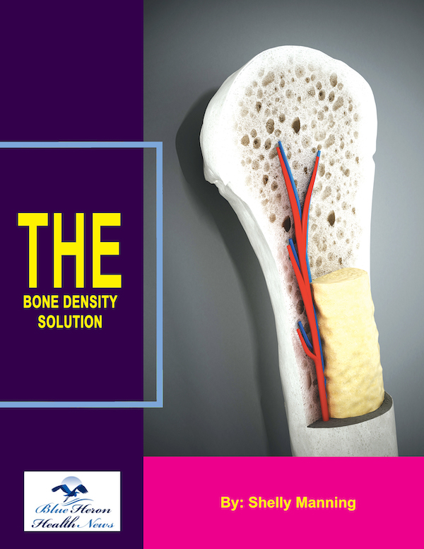
The Bone Density Solution by Shelly Manning As stated earlier, it is an eBook that discusses natural ways to help your osteoporosis. Once you develop this problem, you might find it difficult to lead a normal life due to the inflammation and pain in your body. The disease makes life difficult for many.
How is bone density measured?
Comprehensive Guide to Measuring Bone Density
Bone density, also known as bone mineral density (BMD), is a critical measure of bone health, indicating the strength and risk of fractures. This comprehensive guide explores the various methods used to measure bone density, the technology behind these methods, their applications, and the implications of the results. Understanding how bone density is measured is essential for diagnosing and managing conditions such as osteoporosis.
1. Introduction to Bone Density Measurement
1.1 Definition of Bone Density
- Bone Density: Bone density refers to the amount of mineral matter per square centimeter of bones, indicating their strength and risk of fractures.
- Importance: Measuring bone density helps in diagnosing bone health conditions like osteoporosis, predicting fracture risk, and monitoring treatment effectiveness.
1.2 Purpose of Bone Density Measurement
- Diagnosis: Identifying low bone density and diagnosing conditions such as osteoporosis and osteopenia.
- Risk Assessment: Evaluating the risk of fractures, particularly in older adults.
- Monitoring: Tracking changes in bone density over time to assess the effectiveness of treatments for bone-related conditions.
2. Methods of Measuring Bone Density
2.1 Dual-Energy X-ray Absorptiometry (DEXA or DXA)
2.1.1 Overview
- Gold Standard: DEXA is considered the gold standard for measuring bone density due to its accuracy and reliability.
- Technology: Uses two X-ray beams with different energy levels to measure bone density in the spine, hip, and other areas.
2.1.2 Procedure
- Preparation: Patients lie on a table while a scanner passes over their body. Minimal preparation is required.
- Scan Time: The procedure is quick, usually taking about 10-20 minutes.
- Radiation Exposure: Involves low levels of radiation, comparable to a day’s exposure to natural background radiation.
2.1.3 Interpretation of Results
- T-Score: Compares the patient’s bone density to that of a healthy young adult. A T-score of -1.0 or above is normal, -1.0 to -2.5 indicates osteopenia, and -2.5 or below indicates osteoporosis.
- Z-Score: Compares the patient’s bone density to that of an age-matched population. A Z-score below -2.0 may indicate an underlying problem affecting bone health.
2.2 Quantitative Computed Tomography (QCT)
2.2.1 Overview
- 3D Imaging: QCT provides three-dimensional images and is particularly useful for assessing trabecular (spongy) bone in the spine.
- Accuracy: Offers a more detailed assessment of bone architecture compared to DEXA.
2.2.2 Procedure
- Preparation: Similar to a standard CT scan, requiring patients to lie on a table that moves through a scanner.
- Scan Time: Takes about 10-30 minutes depending on the area being scanned.
- Radiation Exposure: Involves higher radiation exposure than DEXA.
2.2.3 Interpretation of Results
- Volumetric Density: Provides measurements in mg/cm³, offering a detailed analysis of bone density in specific regions.
- Applications: Useful for assessing bone quality and structure, especially in cases where DEXA results are inconclusive.
2.3 Peripheral Quantitative Computed Tomography (pQCT)
2.3.1 Overview
- Peripheral Measurement: pQCT is used to measure bone density in peripheral sites such as the forearm and tibia.
- Application: Helps in assessing bone density in patients who cannot undergo central DXA scans.
2.3.2 Procedure
- Preparation: Patients sit or lie down while the scanner focuses on the specific peripheral site.
- Scan Time: Typically takes 5-10 minutes.
- Radiation Exposure: Lower radiation exposure compared to central QCT.
2.3.3 Interpretation of Results
- Site-Specific Measurements: Provides detailed information about bone density in the scanned peripheral area, useful for monitoring localized bone health.
2.4 Ultrasound Bone Densitometry
2.4.1 Overview
- Non-Invasive Method: Uses sound waves to measure bone density, primarily at the heel (calcaneus).
- Advantages: No radiation exposure and portable equipment.
2.4.2 Procedure
- Preparation: Patients place their foot in an ultrasound device that sends sound waves through the bone.
- Scan Time: The procedure takes a few minutes.
- Application: Often used for initial screening but less accurate than DEXA.
2.4.3 Interpretation of Results
- Quantitative Ultrasound Index (QUI): Measures the speed of sound (SOS) and broadband ultrasound attenuation (BUA) to estimate bone density.
- Risk Assessment: Helps assess fracture risk, especially in settings where DEXA is not available.
2.5 Single-Photon Absorptiometry (SPA) and Dual-Photon Absorptiometry (DPA)
2.5.1 Overview
- Historical Methods: SPA and DPA were early methods of measuring bone density, primarily used before the development of DEXA.
- Technology: SPA uses a single photon beam, while DPA uses dual photon beams for measurement.
2.5.2 Procedure
- Preparation: Similar to DEXA, with patients lying on a table for the scan.
- Scan Time: Takes longer than DEXA, often up to 30 minutes.
- Radiation Exposure: Involves higher radiation exposure compared to modern methods.
2.5.3 Interpretation of Results
- Accuracy: Less accurate and reliable compared to DEXA, thus largely replaced by newer technologies.
3. Applications and Uses of Bone Density Measurement
3.1 Diagnosing Osteoporosis
- T-Score Criteria: Diagnosis based on T-score values from DEXA scans, with a score of -2.5 or below indicating osteoporosis.
- Early Detection: Early diagnosis can help in implementing preventive measures and treatments to reduce fracture risk.
3.2 Assessing Fracture Risk
- Fracture Risk Assessment Tool (FRAX): Combines bone density results with other risk factors to estimate the 10-year probability of fractures.
- Clinical Use: Helps guide treatment decisions based on individual fracture risk.
3.3 Monitoring Treatment Effectiveness
- Follow-Up Scans: Regular bone density measurements to monitor changes in response to osteoporosis treatments.
- Treatment Adjustments: Adjusting treatment plans based on changes in bone density over time.
3.4 Screening and Preventive Health
- Population Screening: Identifying individuals at risk for osteoporosis, especially postmenopausal women and older adults.
- Preventive Measures: Implementing lifestyle changes, dietary modifications, and medications to maintain or improve bone density.
4. Factors Influencing Bone Density Results
4.1 Biological Factors
- Age: Bone density decreases with age, particularly after menopause in women.
- Sex: Women generally have lower bone density than men.
- Genetics: Family history and genetic predisposition play a significant role in bone density.
4.2 Lifestyle Factors
- Diet: Calcium and vitamin D intake are crucial for maintaining bone density.
- Exercise: Weight-bearing and resistance exercises help improve bone density.
- Smoking and Alcohol: Both smoking and excessive alcohol consumption negatively impact bone density.
4.3 Medical Conditions
- Chronic Diseases: Conditions such as rheumatoid arthritis, hyperthyroidism, and chronic kidney disease can affect bone density.
- Medications: Long-term use of corticosteroids, anticonvulsants, and certain cancer treatments can lead to bone loss.
5. Interpretation and Clinical Implications
5.1 Understanding T-Scores and Z-Scores
- T-Score: Comparison to a young, healthy reference population; used for diagnosing osteoporosis and assessing fracture risk.
- Z-Score: Comparison to an age-matched reference population; used to identify potential secondary causes of bone loss.
5.2 Clinical Decision-Making
- Diagnosis and Treatment: Using bone density results to diagnose osteoporosis and guide treatment decisions.
- Risk Management: Implementing strategies to reduce fracture risk based on individual bone density results and overall health.
5.3 Patient Education and Counseling
- Explaining Results: Helping patients understand their bone density results and the implications for their bone health.
- Lifestyle Recommendations: Advising on diet, exercise, and lifestyle changes to improve or maintain bone density.
6. Advances and Future Directions in Bone Density Measurement
6.1 Emerging Technologies
- High-Resolution Imaging: Development of high-resolution imaging techniques for more detailed assessment of bone structure and quality.
- Non-Invasive Methods: Research into new non-invasive methods for measuring bone density with minimal radiation exposure.
6.2 Genetic and Molecular Research
- Genetic Markers: Identifying genetic markers associated with bone density to better predict risk and personalize treatments.
- Molecular Mechanisms: Understanding the molecular mechanisms underlying bone remodeling and density changes.
6.3 Personalized Medicine
- Tailored Treatments: Developing personalized treatment plans based on individual genetic and molecular profiles.
- Precision Health: Using advanced technologies and data to optimize bone health and prevent osteoporosis.
7. Case Studies and Real-Life Applications
7.1 Clinical Case Studies
- Diagnosis and Management: Examples of patients diagnosed with low bone density and their treatment plans.
- Monitoring and Outcomes: Case studies showing how regular bone density measurements impact treatment outcomes.
7.2 Personal Stories
- Patient Experiences: Real-life stories of individuals living with osteoporosis and their journey to manage the condition.
8. Public Health and Awareness
8.1 Education and Outreach
- Public Awareness Campaigns: Initiatives to educate the public about the importance of bone health and preventive measures.
- Community Programs: Local programs offering bone density screenings, exercise classes, and nutritional guidance.
8.2 Policy and Advocacy
- Healthcare Policies: Advocacy for policies that support bone health research, preventive care, and access to treatment.
- Support Networks: Building support networks for individuals with osteoporosis and other bone density-related conditions.
9. Conclusion
Bone density measurement is a crucial tool for diagnosing and managing bone health conditions such as osteoporosis. With various methods available, from DEXA to QCT and ultrasound, healthcare providers can accurately assess bone density, predict fracture risk, and monitor treatment effectiveness. Understanding the factors influencing bone density and the clinical implications of measurement results helps guide effective management strategies and improve patient outcomes. Advances in technology and research continue to enhance our ability to measure and improve bone density, offering hope for better prevention and treatment of bone-related conditions.
The Bone Density Solution by Shelly Manning As stated earlier, it is an eBook that discusses natural ways to help your osteoporosis. Once you develop this problem, you might find it difficult to lead a normal life due to the inflammation and pain in your body. The disease makes life difficult for many.
